Dental tools are essential part of dental offices and laboratories. Dental instruments help dentists, dental hygienists, and dental laboratory technicians in performing dental procedures and fabrication of prostheses. These dental armamentariums are carefully manufactured to meet the precise needs and demands of the dental procedures.
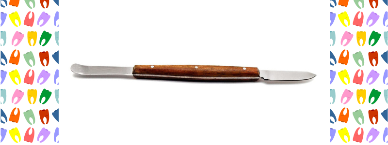
Dental wax carvers are hand-held instruments that are widely used in dental clinical practice and dental laboratories. They are used for carving, shaping, contouring, and sculpting different waxes that help in the fabrication of dental prostheses such as dentures, crowns, and bridges.
Dental wax carvers are typically made of high-quality corrosion-resistant stainless steel. Some dental wax carvers also contain titanium or titanium nitride coatings. The addition of titanium allows the carver to withstand high temperatures and prevent deformation.
Uses of Dental Wax Carvers
The dental use of the Lecron Carveris craving and shaping dental waxes in the dental laboratory and chairside during denture fabrication and the fabrication of other dental prostheses. They are also important tools used during tooth-carving exercises.
They can be used for other purposes, such as carving and sculpting plaster, wax clay, or resins.
Parts of Dental Wax Carver
Dental wax cravers consist of two parts.
Blade: The blade is the working end of the instrument, which comes in contact with wax.
Handle. The operator uses the handle to hold and control the instrument.
Safety precautions
Dental wax carvers are safe and easy to use. However, they still require safety measures and precautions like any other dental instrument. Always wear protective gloves, protective eyewear, and a mask to avoid any accidents during use. While heating the carver do it gently and gradually to avoid overheating of the instrument.
Maintenance and care
Proper cleaning and sterilization of dental wax carvers is important to ensure the longevity and durability of the instrument. Carvers should always be cleaned or sterilized using a disinfectant solution, an ultrasonic cleaner, or an autoclave. Store the carvers in a cool and dry place away from direct heat and moisture to prevent corrosion, tarnishing, and rusting of the instrument. During use the working end of the carver should not be overheated as it will cause deformation and breakdown of the working end of the instrument.
Types of Dental Wax Carvers
Besides Lecron carver there are other different types of wax cravers depending upon the shape of the blade.
PK Thomas: It has a pointed and tapered blade to crave finer details and grooves in the dental wax.
Chisel: straight and sharp blade to carve flat and smooth materials such as waxes and clay.
Scoop: Scoop-shaped curved is used to crave waxes and resins. The scoop-shaped wax carvers are available in different sizes and this type of wax carver can be used with or without heating the wax carver.
Colors and design variations
There are many variations in the design and colors of the wax cravers.
Single-ended carvers have only one blade at the end of the handle.
Double-ended carvers offer more versatility because of blades are present at both ends of the handle.
Colored lecron carvers have single or multicolored tips and handles.
Other Similar Dental Instruments
The dental instruments look similar to the wax carver and are used along with the wax carver are:
In dental school, students are expected to understand the concepts but at times students are expected to memorize a lot of information. Recently, I came across Rememberology dental mnemonics website that can help students to remember information in an easy and fun way. This website is the concept of Dr. Hiba a dental education enthusiast. She would like to make dental concepts and knowledge easily digestible for dental students and professionals.
What Subjects Are Offered by Rememberology Dental Mnemonics Website?
The Rememberology website currently offers dental mnemonics videos for the following subjects.
Oral Pathology:
Two oral pathology courses are available on the website. The first course is the general oral pathology course and the second course is specially designed for the students preparing for the Integrated National Board Dental Examination (INBDE). These courses cover oral hereditary and developmental conditions, defects of the human teeth, salivary gland pathologies, Bone lesions, tumors and cysts, premalignant and malignant lesions, and oral mucosal pathologies.
These courses include short video lectures explaining pathologies using mnemonics, clinical images, histological slides, and radiographs. In addition to that downloadable summary files are available explaining oral pathologies with images, and treatments. These summary files cover almost every pathology of the oral cavity.
Pharmacology:
Pharmacology is a ghost subject for a lot of medical and dental students during and even after their school years. This is a very comprehensive course that covers anesthesia, Antibiotics, Anti-fungal drugs, Antihistamines, drugs related to the cardiovascular system, Analgesics, anti-inflammatory drugs, antidepressants, and mouthwashes.
The course includes short video lectures that explain the classification, mechanism of action, and side effects of drugs in a simple concise way using mnemonics. The mnemonics that are included in this course are especially helpful for the students preparing for Integrated National Board Dental Examination (INBDE), The National Dental Examining Board Assessment of fundamental knowledge (NDEB- AFK). This course also covers the famous NBDE Tufts Pharmacology Board review paper.
At the end of each topic, numerous dental board-style multiple-choice questions are given with amazing explanations. The pharmacology course also includes a downloadable pdf file of 331 pages that includes pharmacology mnemonics and questions.
Head and Neck Anatomy:
Head and neck anatomy is the most important and challenging part for dental school students. The anatomy of the head and neck is an important part of all dental board and dental hygiene board exams.
This course provides some great mnemonics for the anatomy of the head and neck, bones of the skull, the foramina in the skull and the structures that pass through them, and some of the important nerves that supply the head and neck regions.
All of these are explained using pictures, illustrations, and mnemonics in a very interesting way using day-to-day examples.
Oral and Maxillofacial surgery:
The course explains topics that are based on famous dental decks. The course covers syndromes of maxillofacial importance such as Cushing syndrome, Steven Johnson syndrome, nerve and blood supply of the head and neck structures, anesthesia and its classification, indications, and contraindications, medicines related to maxillofacial surgery, bone grafts, and dental implants.
There is a downloadable file that has the same mnemonics as the videos. This pdf file is good for dental students who like review papers.
Periodontology
The periodontology course is based on dental decks and it covers topics such as periodontal surgery, periodontal materials, periodontal diseases such as gingivitis and periodontitis, periodontal diseases during pregnancy, etiology of the inflammation around the dental implant, and management of periodontal disease using local and systemic measures.
The course also offers a concise downloadable file that has the same mnemonics as the videos, and it is good for quick review purposes.
Prosthodontics
The prosthodontics course covers complete denture anatomical landmarks and related clinical aspects, removable and fixed partial dentures, and a brief account of the dental materials related to prosthodontics.
The course also has a downloadable file that provides the same mnemonics as the video related to the applied anatomical aspects of complete dentures and dental materials. This downloadable file is good for a quick review of the course.
Orthodontics
The orthodontics course is a concise course that provides videos with mnemonics on the topic such as tooth size, ectopic tooth eruption, radiographic means to identify the pre-pubertal growth spurt, malocclusion and its types, cephalometric analysis, and orthodontic appliances.
Dental Radiology:
This course covers topics such as radiation exposure and safety for patients and operators, traditional x-ray processing chemicals, errors or artifacts that arise in the film because of processing errors, different maxillofacial views, and their ideal indications (Towne’s view and Water’s view). All videos in the dental radiology course are concise and are explained using mnemonics and illustrations.
Rememberology not only provides you with courses but it also allows you to keep a record and see your progress and achievements through a dedicated dashboard. For an idea of how well and concisely the concepts are explained in each course watch this free preview on Actinomycosis.
Who can get benefits from Dental Rememberology Website?
The Rememberology website is highly recommended for:
Undergraduate and postgraduate dental students.
Dental hygiene students.
Dental nursing students.
Students preparing for dental board exams such as the Integrated National Board Dental Examination (INBDE), The National Dental Examining Board Assessment of fundamental knowledge (NDEB- AFK), the Australian dental council (ADC) exam, and students attempting the National board dental hygiene examination (NBDHE) or other state-specific dental hygiene license exams.
Students currently studying in pre-dentistry programs offered by US universities and intending to enroll in dental school can get a lot of benefits from this website.
Dental faculty can use this website and can make their lectures stimulating for the students.
Medical students can also get benefit from the website as well as the website offering modules such as Pharmacology, Oral Pathology, and Head and Neck Anatomy.
What student’s are saying about the Rememberology website?
Rememberology website is well received by dental students from all around the world. Few of the reviews are
“I would recommend Rememberology not only to NBDE aspirants but also to students/ working dentists to reinforce what they already know.“ (Archie McGill)
“ I love Rememberology ! It just makes studying easier” (Rania)
“Thanks a lot, Dr. Hiba for making all this so easy and fun for us all”. For more student reviews and recommendations visittheRememberology testimonial page.
How much does Rememberology Cost you?
Rememberology offers free previews in each course that will help you in your purchase decision. The 12-month access to all the courses is just 79 US Dollars (USD). Which cost you around 6.5 USD a month which is equal to two Starbucks drinks a month.
The primary mandibular second molar morphologically is similar to the permanent mandibular first molar but it is smaller in all dimensions. This tooth along with the other deciduous molars grind food into smaller particles.
Timeline of Development and Eruption
Following is the summary of the development and eruption of the second deciduous mandibular molar.
Initiation of tooth calcification: 18 weeks in utero
Completion of the crown: 10 months
Eruption of tooth: 27 months
Completion of root: 3 years
The deciduous mandibular second molar is replaced by the mandibular second premolar by the age of 11 to 12 years.
Buccal Aspect
Three cusps are visible from the buccal aspect and these cusps are nearly equal in size.
The cusps are named mesiobuccal, buccal, and distobuccal cusps.
In between the buccal cusps, there are two shallow developmental grooves called the mesiobuccal and distobuccal developmental grooves.
The root trunk of the deciduous mandibular second molar is very small as it divides just below the cementoenamel junction into two roots.
The two roots are the mesial and distal roots. The roots of this tooth are slender, long, and divergent.
Lingual Aspect
There are two cusps on the lingual aspect which are of almost equal dimensions.
The two cusps are named mesiolingual and distolingual cusps.
In between the two lingual cusps a short lingual developmental groove is present.
The cervical line is nearly straight on the lingual aspect.
Parts of the three buccal cusps are visible from the lingual aspect.
Mesial Aspect
On the buccal surface of the crown, there is a prominence near the cervical third portion of the crown.
The mesiolingual cusp is higher as compared to the mesiobuccal cusp.
The cervical line is regular and nearly straight.
The mesial root is broad buccolingually and flat with a blunt root apex.
Distal Aspect
The buccolingual width of the crown is less on the distal aspect.
The distolingual cusp is well developed as compared to the distobuccal cusp.
The distal marginal ridge is shorter and presents slightly at a lower level as compared to the mesial marginal ridge.
The cervical line of the crown is straight buccolingually.
The buccolingual width of the root is less on the distal root, especially in the apical third portion. Therefore, part of the mesial root is visible from the distal aspect.
Occlusal Aspect
All five cusps are visible from the occlusal aspect.
There are three cusps on the buccal side mesiobuccal, distobuccal, and distal cusps.
There are two lingual cusps mesiolingual and distolingual cusps.
The crown tapers towards the lingual side. Therefore, the mesiodistal dimensions are more on the lingual aspect as compared to the buccal aspect.
This tooth appears similar to the permanent mandibular first molar but the overall dimensions of the deciduous mandibular second molar are less. The cusps of the molar are also equal in size, unlike the permanent mandibular first molar.
The central developmental groove is a major developmental groove from which other developmental grooves emerge.
Two triangular fossae are present on the occlusal surface adjacent to the marginal ridges. The mesial triangular fossa is larger as compared to the distal triangular fossa.
Video lecture explaining the morphology of deciduous mandibular second molar
The deciduous mandibular first molar along with the other deciduous molars of the maxillary and the mandibular dental arch grind food into small particles. This tooth does not resemble morphologically with any other deciduous or permanent teeth.
Timeline of Development and Eruption
Following is the summary of the timeline of the development of the deciduous mandibular first molar.
Beginning of calcification: 15.5 weeks in utero
Crown completion: 5.5 months after birth
Tooth eruption: 16 months
Completion of root: 2.5 years
The deciduous mandibular first molar is replaced with the mandibular first premolar around the age of 10-11 years by the process of exfoliation.
Buccal Aspect
The mesial outline of the crown of the deciduous mandibular first molar is almost straight. While the distal outline of the crown it converges from the contact area to the cervical area of the crown.
The height of the distal part of the crown is less compared to the mesial part of the crown.
There are two cusps on the buccal aspect mesiobuccal cusp and the distobuccal cusp.
The mesiobuccal cusp is slightly wider and higher as compared to the distobuccal cusp which is round and smaller.
In between the two buccal cusps a shallow developmental groove is present called the buccal developmental groove.
There are two roots, the mesial and distal roots.
The root trunk on all the deciduous molars including the deciduous mandibular first molar is small and the roots are long and slender.
In the apical third portion, the roots spread beyond the crown outline, especially the distal root.
Lingual Aspect
The crown and the root converge towards the lingual aspect.
There are two cusps on the lingual aspect, mesiolingual and distolingual cusps.
The mesiolingual cusp is long and sharp while the distolingual cusp is short and round.
Parts of the mesiobuccal and mesiolingual cusps are visible from the lingual aspect.
The cervical line is straight on the lingual aspect.
Mesial Aspect
The buccal surface of the tooth on the cervical one-third of the crown has a bulge or convexity.
Mesiobuccal and mesiolingual cusps are visible from the mesial aspect.
A well-developed mesial marginal ridge connects the mesiobuccal cusp with the mesiolingual cusp.
The lingual outline of the crown extends beyond the outline of the root. While the buccal outline is nearly in line with the buccal root outline.
The mesial root is broad buccolingually and it has a square outline. Sometimes a developmental depression extends the full length of the root surface.
Distal Aspect
The cervical line extends almost straight buccolingually on the distal aspect.
The distobuccal and distolingual cusps are not long or as sharp as the mesial cusps. Therefore, parts of the mesiobuccal and mesiolingual cusps are visible from the distal aspect.
The distal marginal ridge connects the distobuccal cusp with the distolingual cusp and it is not well developed.
The distal root is more rounded, shorter, and tapered as compared to the mesial root. Therefore, part of the mesial root is visible from the distal aspect.
Occlusal Aspect
The outline of the deciduous mandibular first molar is irregular.
On the buccal aspect of the crown, prominence is present on the mesiobuccal part.
The mesiolingual cusp is the largest and most well-developed cusp.
The central developmental groove extends from the pit of the mesial triangular fossa and crosses the occlusal surface and terminates into the distal triangular fossa. This central developmental groove divides the occlusal surface into buccal and lingual halves.
There are three fossae on the occlusal surface. The central fossa, mesial triangular fossa, and distal triangular fossa.
The mesial profile of the crown appears flat while the distal profile is convex.
There is numerous supplementary groove that emerges from the main developmental groove.
Video Lecture explaining the morphology of the deciduous mandibular first molar
The overall morphology of the deciduous maxillary second molar is similar to the permanent maxillary first molar. Although the small size, whiteness, short root trunk, and wide divergent roots differentiate the deciduous maxillary second molar from the permanent maxillary first molar.
Development and Eruption
Following is the summary of the development and eruption of the deciduous maxillary second molar.
First evidence of calcification: 19 weeks in utero
Completion of enamel: 11 months
Tooth eruption: 29 months
Root Completion: 3 years
Exfoliation: 10-11 years
The deciduous maxillary second molar is replaced by the maxillary second premolar.
Buccal Aspect
The maxillary deciduous second molar has two well-developed buccal cusps. The cusps are equal in length and development.
The names of two buccal cusps are the mesiobuccal cusp and the distobuccal cusp.
A shallow buccal developmental groove is present between the two buccal cusps.
The crown of the deciduous maxillary second premolar is much larger as compared to the deciduous maxillary first molar.
The root trunk of this root is also short and the point of division of the root is close to the cementoenamel junction.
There are three roots. Mesial, distal, and palatal root.
The roots appear slender, longer, and heavier as compared to the deciduous maxillary first molar.
Lingual Aspect
Three cusps are visible from the lingual aspect. The cusps are the mesiolingual cusp, distolingual cusp, and supplementary cusp.
The supplementary cusp is known as the fifth cusp or tubercle of carabelli.
Mesial Aspect
The crown of the deciduous maxillary second molar has a typical molar outline because of more buccolingual dimensions. The length and development of the root also contribute to the molar-like appearance of this tooth.
Three cusps are visible from this aspect those are the mesiobuccal cusp, mesiolingual cusp, and supplementary or fifth cusp.
The mesial marginal ridge connects the mesiobuccal cusp with the mesiolingual cusp.
Little curvature of the cervical line is evident towards the crown surface.
Distal Aspect
The overall distal calibration of the crown is less as compared to the mesial aspect. Due to this convergence of the crown part of the buccal and palatal aspect of the crown is visible from the distal aspect.
Distobuccal and distolingual cusps are small and are of equal size
The curvature of the cervical line is nearly straight.
All three roots mesial, distal, and palatal roots are visible from the distal aspect. The visibility of all three roots is because the overall dimensions of the distal root are less.
Occlusal Aspect
From the occlusal aspect, all four major cusps (Mesiobuccal cusp, Mesiolingual cusp, Distobuccal cusp, and Distolingual cusp) and one supplementary cusp are visible.
The central fossa is the major fossa on the occlusal aspect of the tooth. The center of the fossa contains the central pit.
Adjacent to the mesial marginal ridge and distal marginal ridges there are two minor fossae called mesial and distal triangular fossae.
A well-defined developmental groove called the central developmental groove crosses the occlusal surface of the tooth.
From the central developmental groove, the buccal groove originates that serves as a demarcation between mesiobuccal and distobuccal cusps. Similarly, the lingual developmental groove originates from the central developmental groove and serves as a demarcation between mesiolingual and distolingual cusps.
Supplementary grooves often originate from the main developmental groove.
A prominent oblique ridge is present that connects the mesiolingual cusp with the distobuccal cusp.
Video Lecture explaining the morphology of the Deciduous Maxillary Second Molar
The deciduous maxillary first molar is smaller than the deciduous maxillary second molar. According to the shape, this tooth is an intermediate between a premolar and a molar. Together with the mandibular molars, the deciduous maxillary molars perform the grinding function.
Development and Eruption
Following is a summary of the development and eruption of the deciduous maxillary first molar.
Initiation of calcification: 15 weeks in utero
Completion of the crown: 6 months
Eruption of tooth: 16 months
Root completion: 2.5 years
Exfoliation of tooth: 10 years
The deciduous maxillary first molar is replaced by the maxillary first premolar.
Buccal Aspect
The buccal surface of the deciduous maxillary first molar is smooth with very few indistinct developmental grooves.
The deciduous maxillary first molar is widest at the contact areas.
There are three roots and all are visible from the buccal aspects. The name of the roots is the mesial root, distal root, and palatal root. The distal root is shorter as compared to the mesial root, and the palatal root is the longest.
The roots of this tooth are slender, long, and widely separated.
The root trunk in the deciduous maxillary first molar is not very well developed. The division of the root begins just below the cementoenamel junction.
Lingual Aspect
The crown converges considerably towards the lingual aspect. Therefore, part of the mesial and the distal aspect of the tooth is visible from the lingual aspect.
The mesiolingual cusp is the most prominent and sharp while the distolingual cusp is smooth and rounded.
The distobuccal cusp is visible because it is long and well developed as compared to the distolingual cusp.
In a three cusp type deciduous maxillary first molar there are two buccal cusps and a large single lingual cusp is present.
All three roots mesial, distal, and palatal are visible from the lingual aspect. The palatal root is the largest.
Mesial Aspect
The mesiolingual cusp of the deciduous maxillary first molar is longer and shaper as compared to the mesiobuccal cusp.
The mesial marginal ridge connects the mesiolingual cusp with the mesiobuccal cusp.
The tooth has a pronounced convexity on the buccal surface at the cervical third of the crown.
The cervical line show curvature towards the occlusal surface.
The lingual root extends lingually. Above one-third of the root the lingual root curves sharply in a buccal direction.
Distal Aspect
The crown of the deciduous maxillary first molar is narrow on the distal aspect as compared to the mesial aspect. Therefore, parts of the buccal and lingual aspects of the tooth are visible from the distal aspect.
The distobuccal cusp is long and sharp as compared to the distolingual cusp which is small and rounded.
Due to small distobuccal and distolingual cusps parts of the mesiobuccal and the mesiolingual cusps are visible from the distal aspect.
The cervical line extends nearly straight from the buccal to the lingual surface of the tooth.
Part of the mesiobuccal root is also visible from the distal aspect.
Occlusal Aspect
The crown of the deciduous maxillary first molar converges towards the lingual aspect.
The crown also converges towards the distal aspect.
The occlusal surface has a central fossa, mesial triangular fossa, and smallest distal triangular fossa.
The central developmental groove extends from the mesial triangular fossa to the distal triangular fossa.
Many supplementary grooves arise from the central developmental groove.
Among the mesial and the distal marginal ridges, the distal marginal ridge is thin and not very well developed.
Video Lecture explaining the morphology of the deciduous maxillary first molar
The primary mandibular canine is smaller in all dimensions. The anatomical landmarks like the cingulum and ridges are less well developed as compared to the maxillary deciduous canine. Along with the maxillary primary canines, the deciduous mandibular canines perform the functions of tearing and holding food.
Development and Eruption
The deciduous mandibular canine emerges into the oral cavity around the age of 20 months. The root of this tooth is completed after three years. The tooth is lost by the process of exfoliation around the age of 9 years and the tooth is replaced by the permanent mandibular canine.
Labial Aspect
The crown and root of the deciduous mandibular canine are shorter in length as compared to the deciduous maxillary canine.
The cusp tip is more towards the mesial side. Due to the mesial shift of the cusp tip the distal cusp slope is larger as compared to the mesial cusp slope.
The cervical ridge is not prominent as compared to the deciduous maxillary canine.
The tooth has a single conical root. In the apical half, there is a slight curvature towards the distal aspect.
Lingual Aspect
The cingulum and the marginal ridges of the deciduous mandibular canine are less developed as compared to the maxillary deciduous canine.
Similar to the marginal ridges the lingual ridge which divides the lingual fossa into two halves is also not very prominent.
Due to less development of the ridges and the cingulum, the lingual surface of the crown is smooth with shallow lingual fossae.
The crown and the root of the deciduous mandibular canine tapers towards the lingual aspect. Therefore, part of the mesial and distal aspects of the crown and the root are visible from the lingual aspect.
Mesial Aspect
The labiolingual dimension of the mandibular deciduous canine is less as compared to the maxillary deciduous canine.
The cervical ridges that are present on the labial and the lingual portion of the crown are less prominent as compared to the deciduous maxillary canine.
The mesial root surface is smooth with no developmental depression.
Distal Aspect
The cervical line curvature is towards the incisal aspect. The extent of the curvature of the cervical line on the distal aspect is less as compared to the mesial aspect.
The root surface on the distal aspect has a shallow developmental depression.
Incisal Aspect
The labiolingual dimension of the tooth is slightly less as compared to the deciduous maxillary canine.
The labial surface is convex at the cervical third of the crown.
The cusp tip is more towards the mesial aspect.
The lingual surface of the tooth from the incisal aspect appears smooth.
Video Lecture Explaining the Morphology of the Primary Mandibular Canine
In the deciduous dental arch deciduous maxillary canine are the third teeth from the midline. The deciduous maxillary and the mandibular canines punch, tear, and apprehend food material.
Development and Eruption
The deciduous maxillary canine emerges into the oral cavity by the age of 19 months. The root of the tooth is completed by the age of 3 years. These teeth are replaced by the permanent maxillary canine by the age of 11 to 12 years.
Labial Aspect
The cusp tip of the deciduous maxillary canine is sharper than permanent maxillary canine.
The labial outline of the crown is a diamond shape.
The mesiodistal dimension of the crown is slightly more as compared to incisocervical length of the crown.
The mesial cusp slope is larger as compared to the distal cusp slope. In the permanent maxillary canine, the mesial cusp slope is shorter as compared to the distal cusp slope.
A labial ridge is present on the crown surface that extends from the cusp tip to the cervical area of the crown.
A single conical root is present that inclines towards the distal aspect.
Lingual Aspect
A lingual ridge is present on the crown of the deciduous maxillary canine that divides the lingual fossa into two fossae.
The fossa that is present near the mesial part is called the mesiopalatal fossa. The fossa that is near the distal part of the crown is the distopalatal fossa.
The mesial and the distal marginal ridges are not well developed as compared to the marginal ridges of other deciduous maxillary anterior teeth and permanent maxillary canine.
The root of the crown is longer as compared to the length of the crown.
The root of the deciduous maxillary canine is long as compared to all deciduous teeth.
Mesial Aspect
The labiopalatal dimension of the tooth is more as compared to the labiopalatal dimension of the deciduous maxillary central and lateral incisors.
The labial cervical ridge is prominent.
The curvature of the cervical line is more and it is towards the crown surface.
Distal Aspect
The curvature of the cervical line is less on the distal aspect as compared to the mesial aspect.
The root of the deciduous maxillary canine appears bulky. The greater bulk of the root is in the cervical and middle third of the crown.
Incisal Aspect
The crown of the deciduous maxillary canine is diamond in shape.
The cusp tip of this tooth is offset more towards the distal side.
The cingulum is usually present in the center on the lingual aspect but sometimes slightly offset towards the distal aspect.
The lingual ridge divides the palatal fossa into mesiopalatal and the distopalatal fossae.
Video Lecture Explaining the Morphology of the Primary Maxillary Canine
The deciduous mandibular lateral incisor is wider than the deciduous mandibular central incisor and asymmetrical. The deciduous incisors have a common function which is cutting food. The morphology of the tooth is similar to its permanent successor which is permanent mandibular lateral incisor.
Development and Eruption of Deciduous Mandibular Lateral Incisor
The calcification of the deciduous mandibular lateral incisor begins around the age of 16th week in utero. The tooth emerges into the oral cavity by the age of 13 months and the root is completed by the age of 1.5 years. The deciduous mandibular lateral incisor is replaced by the permanent mandibular lateral incisor by the age of 7 to 8 years.
Labial Aspect
The labial surface of the crown of the deciduous mandibular lateral incisor is smooth with no developmental depression or groove.
The Crown is wider and longer than the deciduous mandibular central incisor. A similar pattern is observed in the permanent mandibular incisors.
The incisal ridge of the deciduous mandibular lateral incisor has a slope towards the distal side.
The distoincisal angle of the tooth is more rounded as compared to the mesioincisal angle.
In the apical third the root has a slight curvature towards the distal side of the tooth.
The root of this tooth is longer than the root of the deciduous mandibular central incisor.
Lingual Aspect
The cingulum of the primary mandibular lateral incisor is more well developed as compared to the cingulum of the deciduous mandibular central incisor.
The mesial and the distal marginal ridges are also more well developed as compared to the deciduous mandibular central incisor.
Due to well-developed marginal ridges and cingulum, the lingual fossa is slightly deeper in comparison with the deciduous mandibular central incisor.
The mesial and the distal aspect of the crown and the root are slightly visible because of the crown and the root convergence towards the lingual aspect.
Mesial Aspect
Convexity at the cervical third of the crown on the buccal and lingual aspect is pronounced.
The mesial surface of the root of the deciduous mandibular lateral incisor is flat and evenly tapered towards the apex of the root.
The cervical line of the tooth has a curvature towards the incisal aspect of the crown.
Distal Aspect
The cervical line on the distal aspect of the deciduous mandibular lateral incisor is less curved towards the incisal aspect.
A longitudinal developmental groove or depression is present on the root surface.
Incisal Aspect
The crown of the deciduous mandibular lateral incisor is not symmetrical as compared to the deciduous mandibular central incisor which is symmetrical.
This asymmetry in shape is because of the curvature of the incisal ridge and position of the cingulum which is offset more towards the distal side.
The labial surface of the crown is more convex as compared to the deciduous mandibular central incisor.
The center of the lingual surface of the crown is more concave because of well-developed marginal ridges and cingulum.
Video Lecture of the Morphology of Deciduous Mandibular Lateral Incisor